Home
/ Lower Leg Bones Diagram / fibula | bone | Anatomy bones, Medical anatomy, Physiology : Bones of lower leg and foot diagram lower leg compartments.
Lower Leg Bones Diagram / fibula | bone | Anatomy bones, Medical anatomy, Physiology : Bones of lower leg and foot diagram lower leg compartments.
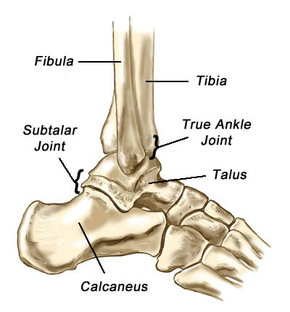
Lower Leg Bones Diagram / fibula | bone | Anatomy bones, Medical anatomy, Physiology : Bones of lower leg and foot diagram lower leg compartments.. The two bones beneath your knee that make up your shin are your tibia and fibula. The human skeleton bones structure function teachpe com, seer training classification of bones, lower leg bones 1024 x 1350 anatomy system human body, human skeleton long femur bone diagram get rid of wiring diagram problem. The foot bones shown in this diagram are the talus, navicular, cuneiform, cuboid, metatarsals and calcaneus. At the microscopic level, this hard outer. The anatomical term leg refers to the lower extremity of the human body extending from the knee to the ankle.
(2) hip bone attaches legs to our body. Your leg bones are the longest and strongest bones in your body. The leg is the region of the lower limb between the knee and the foot. Muscles of the leg and foot classic human anatomy in motion: Bone chart insaat mcpgroup co.
Muscles of the Lower Extremities. Muscular system from encyclopedia.lubopitko-bg.com Click now to learn more about the bones, muscles, and soft tissues of these regions at kenhub! The forearm and the lower leg have two long bones each. At the distal end of the femur, two rounded condyles meet the tibia and fibula bones of the lower leg to form the knee joint. Calcaneus, talus, navicular medial cuneiform, intermediate cuneiform, lateral cuneiform and cuboid. Lower bones limbs limb leg diagram muscle foot template anatomy blank human skeleton coloring sketch function th. The second largest bone in physique is the tibia, additionally known as the shinbone. Anterior view with primary bones names. Vtt 150 horse leg anatomy diagram quizlet.
Continue scrolling to read more below.
Continue scrolling to read more below. The human leg, in the general word sense, is the entire lower limb of the human body, including the foot, thigh and even the hip or gluteal region. The knee joint is the largest joint in the body and is primarily a hinge joint, although some sliding and rotation occur. The upper leg bone is connected to the lower leg bones at the knee by a hinge joint. The human leg consists of 8 bones, 4 per leg. Anterior view with primary bones names. Bones of lower leg and foot diagram lower leg compartments. What is the weight bearing bone of the lower leg? Your upper and lower leg are connected by a hinge joint. Cheek bone (zygoma) upper jaw (maxilla). Vector illustration with human skeleton scheme vector illustration anatomy of human legs and diagram of human bones isolated on white background. The tibia and the fibula. Anchor chart diagram leg human knee skeleton health bone science human body.
The two bones beneath your knee that make up your shin are your tibia and fibula. Download a free preview or high quality adobe illustrator ai, eps, pdf and high resolution jpeg versions. The femur, or thigh bone, is the largest, heaviest, and strongest bone in the human body. It is sometimes called the lower leg. The artist's guide to the.
Ankle & Lower Leg Anatomy - Foot, Ankle & Lower Leg ... from leassessment.weebly.com He leg's main function in the human is for locomotion and support of the rest of the body. Calcaneus, talus, navicular medial cuneiform, intermediate cuneiform, lateral cuneiform and cuboid. It is sometimes called the lower leg. The forearm and the lower leg have two long bones each. Human skeleton parts functions diagram facts britannica. Lower leg muscle diagram blank sketch coloring page. It is the tibial joint surface or ceiling of the ankle mortise. Your leg bones are the longest and strongest bones in your body.
8 4 bones of the lower limb anatomy and physiology.
The lower limb (excluding the foot and image: When you stand or walk, all the weight of your upper body rests on them. The foot bones shown in this diagram are the talus, navicular, cuneiform, cuboid, metatarsals and calcaneus. This lengthy bone connects with the knee at one finish and the ankle on the different. At the distal end of the femur, two rounded condyles meet the tibia and fibula bones of the lower leg to form the knee joint. Vector illustration with human skeleton scheme isolated on a white background. By natalia kremenon january 21, 2021in wiring diagram231 views. Blank bone diagram rome fontanacountryinn com, low satisfaction fishbone free low satisfaction fishbone templates, long bone diagram timothyakeller flickr bone diagram barca fontanacountryinn com. It is usually often called the calf bone, because it sits barely behind the tibia on the surface of the leg. These simple labelled diagrams of the bones of the lower legs and feet and the bones of the arms and hands this diagram shows the skeletal structure of the leg (anterior view) and foot (dorsal view). The bones involved in it, however, are only the femur and the tibia, although the smaller bone of the leg, the fibula, is carried along in the movements of flexion, extension, and slight rotation that this joint permits. He leg's main function in the human is for locomotion and support of the rest of the body. The shoulder is made up of three bones:
Bones of lower leg and foot diagram lower leg compartments. Download a free preview or high quality adobe illustrator ai, eps, pdf and high resolution jpeg versions. Related posts of bone anatomy lower leg. The upper leg bone is connected to the lower leg bones at the knee by a hinge joint. However, in the world of anatomy, the 'leg' strictly means.
leg bones - DriverLayer Search Engine from encyclopedia.lubopitko-bg.com The tibia (shin bone) is the medial bone of the leg and is larger than the fibula, with which it is paired (figure 3). Cheek bone (zygoma) upper jaw (maxilla). The forearm and the lower leg have two long bones each. Muscles of the leg and foot classic human anatomy in motion: 8 4 bones of the lower limb anatomy and physiology. A diagram showing the main bones of the leg and knee. Describe the bones and bony landmarks that articulate at each joint of the lower limb. Posted on january 21, 2015 by admin.
Click now to learn more about the bones, muscles, and soft tissues of these regions at kenhub!
However, in the world of anatomy, the 'leg' strictly means. The foot bones shown in this diagram are the talus, navicular, cuneiform, cuboid, metatarsals and calcaneus. Leg muscle diagrams these pictures of this page are about:lower leg bones diagram. Name the 7 bones of the foot (not counting the phalanges). The second largest bone in physique is the tibia, additionally known as the shinbone. Knee human anatomy function parts conditions treatments. Anterior view with primary bones names. Muscles of the leg and foot classic human anatomy in motion: The human leg consists of 8 bones, 4 per leg. At the distal end of the femur, two rounded condyles meet the tibia and fibula bones of the lower leg to form the knee joint. However, the definition in human anatomy refers only to the section of the lower limb extending from the knee to the ankle, also known as the crus. The lower limb (excluding the foot and image: The human leg, in the general word sense, is the entire lower limb of the human body, including the foot, thigh and even the hip or gluteal region.
What is the weight bearing bone of the lower leg? leg bones diagram. A diagram showing the main bones of the leg and knee.

